 This Web page has been archived on the Web.
Archived Content
Information identified as archived on the Web is for reference, research or recordkeeping purposes. It has not been altered or updated after the date of archiving. Web pages that are archived on the Web are not subject to the Government of Canada Web Standards. As per the Communications Policy of the Government of Canada, you can request alternate formats on the Contact Us page.
Tomentosus Root Rot
Inonotus tomentosus (Fr.:Fr.) S. Teng.
(= Polyporus tomentosus (Fr.:Fr.)
(=Onnia tomentosa (Fr.) P. Karst)
Basidiomycotina, Aphyllophorales, Polyporaceae
Hosts:Inonotus tomentosus has been reported in B.C. on amabilis and subalpine fir, Engelmann, black, and white spruce, lodgepole, ponderosa, and white bark pine, Douglas-fir, western hemlock, and western larch. In other parts of North America it has also been found on grand fir, western larch, western white pine, Sitka spruce, and western redcedar.
Distribution:Inonotus tomentosus is found most frequently in the spruce-pine forests in central and northern British Columbia, and at higher elevations in southern B.C.
Identification: Advanced root infection by I. tomentosus can often be recognized by reduced leader and branch growth, thinning of the foliage (Figs. 3a, 3b), stress cone crops, and death of the tree. Although rare, cankers and resinosus may be present at the base of stems and near the root collar. Wind-thrown trees or groups of dead and dying trees may indicate the presence of the disease in a stand. Fruiting bodies of I. tomentosus are small, usually less than 10 cm in diameter, stalked, and are found on the ground around infected trees (Fig. 3c). Similar, shelf-like fruiting bodies are also found on dead roots and at the base of infected stems (Fig. 3d), but these are generally produced only by a related species, I. circinatus. Fruiting bodies are annual and leathery, most commonly developing in August and September. The upper surface is yellow-brown to rust-brown and velvety. The whole fruiting body becomes dark brown with age.
The early stage of the decay is characterized by a red-brown discoloration in the heartwood (Fig. 3e). The advanced decay has large, elongated to rectangular spindle-shaped pits, separated by red-brown firm wood (Figs. 3f, 3g, 3h). The cross section of an infected stem has a honeycomb appearance. Stump surfaces often demonstrate these stages of decay and can be used to identify the presence of the disease (Figs. 3i, 3j, 3k).
Microscopic Characteristics: Contextual hyphae simple septate. Basidiospores hyaline, negative in Melzer's reagent, acyanophilous, smooth, ellipsoid, 5-6 x 3-4 µm. Setae are abundant, 7-11 x 50-70 (140) µm, straight (Fig. 3l) (compared to I. circinatus, which has hooked setae, Fig. 3m). Growth in culture slow, mat yellow-brown to brown, laccase positive. Stalpers: 1 3 4 (9) (11) (14) (15) 17 (18) 24 25 (26) 28 (30) (31) 34 (35) 38 48 52 53 (54) 64 67 80 [89] 90.
Damage: Infected trees may appear healthy but have extensive butt cull and reduced annual increment growth. The fungus spreads from tree to tree at points of root contact; consequently, diseased trees occur in groups and mortality results in "stand openings." Windthrow may occur before the death of an infected tree. Fruiting bodies on or around a tree indicate that three or more metres of rot may be present in the base of the stem. Inonotus tomentosus is a serious problem in second-growth stands as the stumps of infected trees provide an inoculum source for young trees.
Remarks: Two similar species of Inonotus are found in B.C., I. tomentosus and I. circinatus, which is thought to be less virulent. It is difficult to differentiate between the two, particularly when basidiocarps are not present. Basidiocarps of I. tomentosus are smaller and thinner and are usually form in groups whereas those of I. circinatus are larger, thicker, and tend to be found singly. Inonotus circinatus is less commonly found on spruce than I. tomentosus. Setal characteristics are a good diagnostic feature. The advances stages of decay may also be confused with that of Phellinus pini.
References:
Hunt, R. S. and L. Unger. 1994. Tomentosus root disease. Can. For. Serv., Forest Pest Leaf. No. 77. Victoria, B.C.
Whitney, R. D. 1977. Polyporus tomentosus root rot of conifers. Can. For. Serv. For. Tech. Rep. No. 18.
Figures
Click on any image to see the full size version.
Press "Back" on your browser to return to this screen.
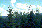
Figure 3a: Healthy (right) and I. tomentosus-infected (left) spruce. The diseased tree shows reduced growth and a distress-crop of cones.
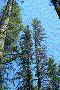 Figure 3b: Crown symptoms in mature diseased spruce.
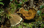 Figure 3c: Fruiting bodies of I tomentosus.
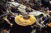 Figure 3d: Fruiting bodies of I circinatus.
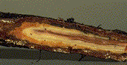 Figure 3e: Heartwood discoloration in an infected root.
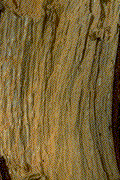 Figure 3f: White pocket rot symptoms.
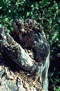 Figure 3g: Honeycomb appearance of an infected root.
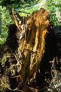 Figure 3h: Honeycomb appearance of an infected root.
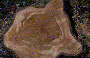 Figure 3i: Decay evident in cross-sections of I. tomentosus-infected spruce stumps.
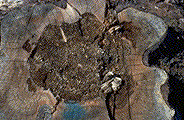 Figure 3j: Decay evident in cross-sections of I. tomentosus-infected spruce stumps.
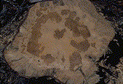 Figure 3k: Decay evident in cross-sections of I. tomentosus-infected spruce stumps.
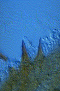 Figure 3l: Setal hyphae of I. tomentosus .
 Figure 3m: Setal hyphae of I. circinatus .